|
|
||||||||||||||||||||||||||||||||||||||||||||||||||
|
Treatment
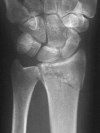
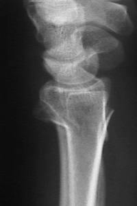
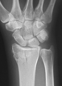
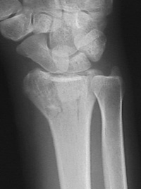
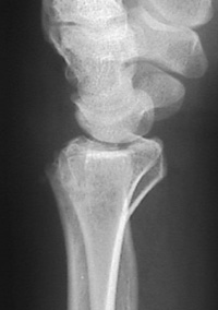
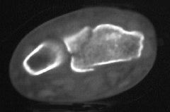
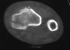
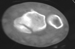
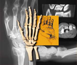
After reviewing the plain radiographs, the patient was sent for CT scans of both wrists.
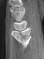
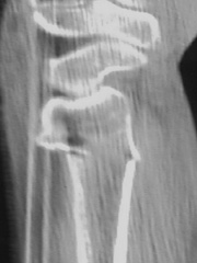
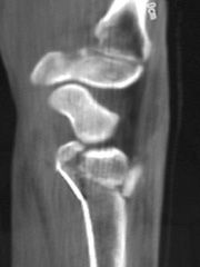
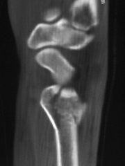
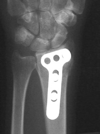
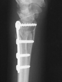
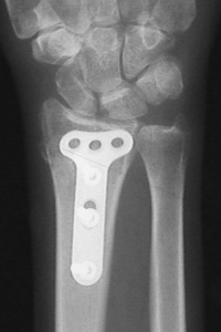
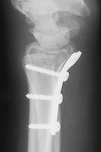
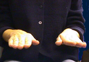
The CT scans showed intraarticular fractures of both distal radii, as well as significant comminution. After discussion with the patient, she had open reduction and internal fixation of both wrists approximately one week apart. The left wrist was operated on first. In the operating room, the fracture was reduced and a dorsal T-plate was applied. The following week, the right distal radius underwent a volar open reduction with placement of a buttress plate. Bone grafting was not performed in either fracture. Post operatively, the regimen was identical for each fracture. Digital motion was begun on the first postoperative day. The initial operative splint was removed two weeks after surgery. The patient was placed in custom molded, removable wrist control splints. She began gentle active motion of the wrist (flexion/extension and pronation/supination) with the therapists three times per week. Six weeks (right wrist) and seven weeks (left wrist) postoperatively, radiographs were taken which revealed fracture healing. The patient was then instructed to wean herself from the splints over the following two to three weeks. At nine and ten weeks after surgery for the right and left wrists, respectively, she had discontinued the use of splints. Her wrist motion on the right is: flex/ext=37/55, pro/sup=75/80; motion on the left is: flex/ext=40/60, pro/sup=70/55. Open reduction and internal fixation was chosen to allow early motion of the wrist. A dorsal plate was chosen for the left wrist because of the displacement of the intraarticular component of the fracture combined with dorsal angulation and metaphyseal comminution. The plate serves as a buttress against dorsal angulation with a single radial styloid screw in the largest of the distal fragments for additional support. The right wrist is a comminuted variant of the chauffeur's
fracture with volar displacement of the articular fragment. The volar
fragments were held together by periosteum, which was left intact. The
plate was used to buttress the volar fragments after reduction.
For discussion of this case, click on Forum (below), then look under Guest Professor, and click on Case 3: Bilateral Fractures.
|
||||||||||||||||||||||||||||||||||||||||||||||||||
| About Us | Research | Basic Knowledge | What's New | Forum | Guest Professor | Post a Case | eRadius Conference | Patients | Home |