David Ring, MD
Fellow, Orthopaedic Hand Service
Massachusetts General Hospital
ACC 527
15 Parkman St.
Boston, MA 02114
(submitted 1999)
(*David is now an attending at MGH)
|
Jesse B. Jupiter, MD
Professor
of Orthopaedic Surgery
Harvard Medical School
Director, Orthopaedic Hand Service
Massachusetts General Hospital
ACC 527
15 Parkman St.
Boston, MA 02114 |
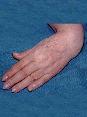
A 77-year-old female employed in clerical work fell from a standing
height fracturing her left, non-dominant distal radius and ulna.
The fracture was treated in a cast, but she continued to have problems
with pain and deformity. She presented to our office 2 years after
the injury complaining of pain and instability of the wrist leading
to severe dysfunction.
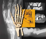
Figure 1: This clinical radiograph demonstrates the severe radial
deviation deformity present. This represents a post-traumatic club
hand deformity. The extreme radial deviation of the wrist has led
us to refer to this deformity as a golf-club hand.
 |
 |
| |
|
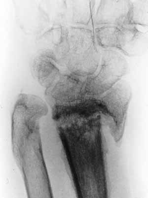
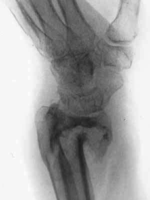
Figure 2 A and B: Radiographs demonstrated
synovial nonunion of the distal radius and ulna. The distal
articular fragment of radius is very small. This is due, in
part, to erosion from the synovial process. |
|
Questions to Consider:
(1) Why didn't the fracture heal?
(2) When considering treatment, what are reasonable goals?
(3) What treatment would you recommend?
(4) If you were to attempt to heal the fracture, how would you secure
the small distal fragment?
The case discussion continues on the next
page>>>
|
|