The Accuracy of Plain Radiographs and CT Scans
in Determining Intra-articular Displacement in Distal Radius Fractures
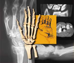
Nelson, D.L., Dowd, J.D., Summers, R.A., and Stoller, D.W.: Accuracy of Plain Radiographs and Computed Tomography Scans in Determining Displacement in Distal Radius Fractures. Presented at the 62nd Annual Meeting of the American Academy of Orthopedics, Orlando, Florida, February 16-21, 1995. A model of a die-punch fracture of the distal radius was created in a cadaver. The amount of the intra-articular step-off was adjustable by means of a screw-driven external fixator and was set at 0, 0.5 mm, 1.0 mm, and 2.0 mm. Plain radiographs and CT scans were obtained and the deformity blindly read by twenty orthopedists and radiologists. Results showed that there was an overestimation of the step-off by 1 mm or more in 8.5% of the plain radiographs and 17% of the CT scans. There was an underestimation of the step-off by 1 mm or more in 36% of the plain radiographs and 21% of the CT scans. The observers were asked to estimate the upper limit of their expected error. They exceeded this limit in 23% of the plain radiographs and in 29% of the CT scans. With this model, the CT scan was not statistically significantly more accurate than the plain radiographs. Both the volar cortex and the central fossa were found to be more reliable as radiographic landmarks on the plain radiograph than the dorsal cortex, but there was no difference between the volar cortex and the central fossa. We found that estimates of step-off cannot be made reliably at the level of 1 mm, and observers were not accurate in their estimation of their upper limit of expected error. In view of our limited ability to accurately determine step-off at a level of 1 mm, the concept of a specific relationship between a given degree of deformity measured in millimeters and outcome must be questioned. |
|