|
|
||||||
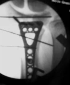
(case submitted August 10, 2007) Synopsis: 35 YO white male fell off his horse playing polo. The patient underwent an ORIF with a volar plate. The bone fragments were difficult to visualize, due to the amount of tissue damage, clot, callus, and torn fat and fascia overlying the pronator quadratus. Visualization was much better after a careful debridement. The shaft and articular comminution were challenging to manage, as it was impossible to reduce all of the fracture lines at once. Therefore, the brachioradialis was released, then articular surface was reduced to itself and pinned next. The articular surface was not reduced to the metaphyseal bone at this time. The major shaft fragment was pinned to the shaft. The metaphyseal bone was left for later.
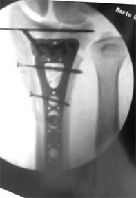
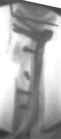
The fracture seemed amenable to volar plating, so an ex fix was not felt to be necessary. The K-wires had been placed to avoid conflict with the plate screws. The plate was applied distally and used to reduce the articular surface to the metaphyseal bone. The alignment looked good, and the shaft K-wires were replaced with 2.4 mm cortical screws. The K-wire in the subchondral bone was left in place.
|
||||||
| About Us | Research | Basic Knowledge | What's New | Forum | Guest Professor | Post a Case | eRadius Conference | Patients | Home |