|
|
||||
|
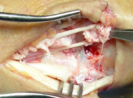
The mass was approached through a transverse incision centered over the midpoint of the third metacarpal. It was found to be a fluid-filled mass enveloping the extensor tendons, which appeared normal. The mass was larger than anticipated, and the incision was extended twice. The mass seemed to track towards the wrist, and finally a "T" extension was made, longitudinally over the wrist, and the fourth extensor compartment opened. The EIP and EDC II tendons were found to be shredded:
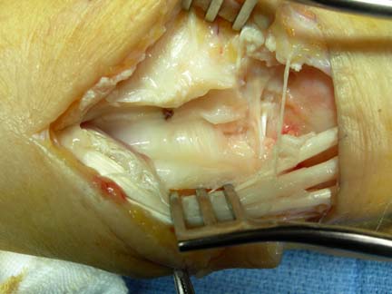
The tendons were moved out of the third and fourth compartment and the floor of the compartments examined. This is what was found:
The tip of one of the volar plate screws was protruding into the floor of the edge of the third and fourth compartments.
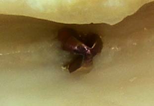
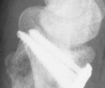
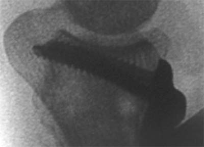
The screw appeared to protrude just slightly less than one mm into the third and fourth compartments, but it was obviously enough to severely injure the tendons. The tendons are in close apposition to the bone in the extensor compartments, with no ability to move out of the way. Over time, the screw tip progressively eroded the tendon. Retrospective review of the lateral radiographs demonstrates the problem:
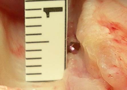
Closeup views of two of the facet laterals, with contrast and brightness digitally optimized. The ulna is long and the facet laterals make it seem longer. It therefore overlies the dorsal aspect of the radius. The screw is seen to be extending over the dorsal surface of the radius. This was not appreciated on any of the views prior to the intraoperative discovery of the protruding screw tip. Had you discovered it during your review of the case? Back on Question 5, had you considered the screw too prominent?
Close examination of the lateral is necessary. Most orthopedic screw placement should have bicortical purchase in order to optimize the stability of the fixation. Past pointing, of about 1 to 2 screw diameters, is recommended, to allow full thread purchase of the far cortex (the self-tapping cutting flutes decrease the purchase at the very tip). However, in the case of volar fixation of the distal radius, the far cortex is the dorsal cortex, which does not offer any significant purchase: it is very thin and often comminuted. The fixation comes from both the thicker volar cortex and the subchondral bone. Examine any lateral xray of the radius and the density of these two areas in comparison to the dorsal cortex is obvious. In addition, any past pointing of the distal screws will endanger the tendons, which are in close apposition to the bone. There is well under 1 mm of periosteum and other soft tissue between the dorsal cortex and the volar aspect of the extensor tendons. In this case, the tendons were 25% intact and were thought to be in no danger of further attritional tearing once the screw was removed. A volar incision was made and the screw removed. The tendon tears were trimmed to give the tendon a smooth surface and the extensor compartment closed with a relaxing incision to decrease the force directing the tendons against the bone. Comments on this case can be made in the Forum. Click on Case #12. |
||||
| About Us | Research | Basic Knowledge | What's New | Forum | Guest Professor | Post a Case | eRadius Conference | Patients | Home |