|
|
||||||||||||||||
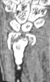
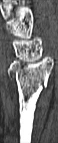
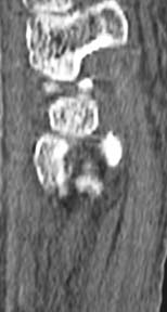
There were two issues. The first issue is the size of the gap. My best estimate of the gap was that it was 4 mm, possibly 3 mm, certainly not 5 mm.
A prior study done in my lab using a cadaveric model of a distal radius fracture has demonstrated that the CT scans of 13 years ago could not accurately measure a gap or stepoff to an accuracy of 1 mm, that plain films were only slightly more accurate, and that hand surgeons are often off in their reading of a CT or plain film by 1 mm yet are absolutely certain that they are not off by more than 1/4 mm. It remains to be seen if the current generation of CT scanners are any more accurate. I am negotiating with the Radiology Department to undertake such a study, but they have shown a decided uninterest in the topic. There is no solid evidentiary basis for using a specific measurement of stepoff or gap as an indication for surgery. Despite the widespread citation of the 1987 Knirk and Jupiter paper on this issue, Dr. Jupiter has repeatedly withdrawn the conclusion that there is a tight correlation between stepoff and the subsequent development of arthritis. He has done this in a prior posting on eRadius and most recently at the 2005 ASSH meeting, in a presentation on "Papers I Wish I could Withdraw." There has been a sizeable amount of papers and presentations on this subject (see Indications for Reduction), but there is no solid evidence for a specific threshold in mm for either stepoff or gap that would indicate that a fracture should be reduced, in part due to the fallibility of measuring gap or stepoff by plain radiographys or CT's. This data void does not relieve the surgeon faced with a specific patient, however. My current indication for surgery, in a reasonably active patient, is I will observe 1 mm of stepoff and attempt to surgically reduce 2 mm of stepoff, and will observe 2 mm of gap and attempt to surgically reduce 3 mm of gap. However, I do not fool myself by thinking that there is a solid base of evidence for this decision; it is more the art than the science of medicine, and I await better studies in the future.
The second issue was the patient's desire to return
to work as soon as possible. I believe that there are two scenarios in which I am currently offering the option of surgery (as opposed to recommending surgery), as an alternative to a six-week period of cast immobilization, for a patient who has an acceptably aligned distal radius fracture. Scenario 1: Working patients or athletically active patients who feel that they would prefer an ORIF to cast immobilization. I think that the risks of ORIF with a volar fixed angle plate are low enough that it is reasonable to offer this option to patients. This probably represents the greatest change in my surgical lifetime in the management of distal radius fractures. Could you, a surgeon, work with a cast? No. Could you support your family for six weeks with a cast? No. I think we need to offer the same consideration to our patients. Scenario 2: Elderly patients, especially if they have DJD and/or live alone, who feel that they would prefer an ORIF to six weeks of cast immobilization. Many elderly patients are significantly incapacitated by a cast. Many would not drive, which makes them isolated and has a negative effect on their ability to eat properly. Many would not wash dishes with a cast and therefore would stop cooking in large part. Many with DJD will develop significant stiffness in their shoulder, elbow, and wrist, despite our interventions. Their activities of daily living, from simple dressing to self care to perineal hygiene, would be strongly effected. I now offer an ORIF to them as an alternative to a cast, outline the advantages and disadvantages of each option, and let them decide. I do not try to slant the discussion one way or the other. I have presented these two scenarios at multiple national and international forums over the last year, and have been surprised at the number of surgeons who have adopted a similar treatment paradigm over the last few years. It is important to not overlook the radial nature of this paradigm shift: it is the most radical change in the management of distal radius fractures in my surgical lifetime. My treatment: on the next page >>> |
||||||||||||||||
| About Us | Research | Basic Knowledge | What's New | Forum | Guest Professor | Post a Case | eRadius Conference | Patients | Home |